
Comprehensive Care - 1a
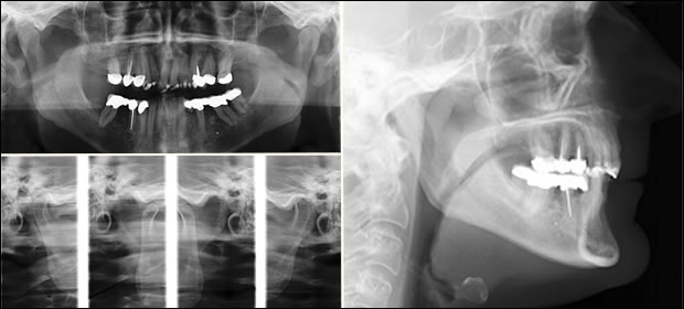
Fig 6: Diagnostic images for this case included a panoramic image, trancranial images of the temporo-mandibular joints in the closed and open positions, as well as a cephlometric image.


Fig 7: Occlusal views demonstrate the defective restorations, enamel abrasion, and crooked teeth. It was decided that treatment would be restricted to the anterior teeth. A multi-disciplinary approach was essential in achieving a successful functional and esthetic result.
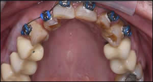
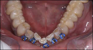
Fig 8: The maxillary anterior teeth were slenderized and orthodontic appliances were inserted.
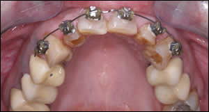
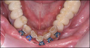
Fig 9: Nickel titanium wires with Tip-Edge brackets provided rapid leveling and unraveling of the incisors with light force.
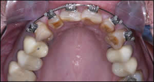
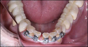
Fig 10: A coil spring was used to position tooth # 7 equidistant between tooth #6 and 8. An elastic chain was utilized on the lower arch to close the space that was created by uprighting tooth #24 and 26. Note tooth #25 is missing.
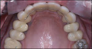
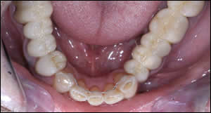
Fig 11: Total treatment time was 3 months for the maxillary teeth. Splinted interim acrylic crowns were placed on the maxillary anterior teeth to achieve golden proportions...and to provide retention. Treatment time was 6 months for the mandibular teeth. The alignment of the mandibular anterior teeth allowed for the proper contour and sagital position of the upper incisors. A normal overbite/overjet relationship was established. A bonded lingual retainer was placed on the lower anterior teeth.